Urokinase-type Plasminogen Activator [u-PA] Assays
Introduction
Urokinase-type Plasminogen Activator or u-PA is synthesised as a single chain glycoprotein [Pro-uPA] consisting of 411 amino acids. Following secretion, u-PA is cleaved [Lys158-Ile159] to generate a significantly more active two-chain form linked by a disulphide bond. A number of proteases can potentially cleave the single chain u-PA to the two chain u-PA including Plasmin. This molecule can be further processed into a catalytically active low molecular weight Urokinase-type plasminogen activator (LMW-uPA).
u-PA is involved in the conversion of Plasminogen to Plasmin and the regulation of Fibrinolysis. Although originally identified in the urine and hence its name, it is present in a number of other tissues.
u-PA binds to the u-PA receptor [u-PAR] which localises u-PA to the cell surface. u-PAR can also binds to integrins and other cell surface receptors to activate different intracellular signalling pathways.
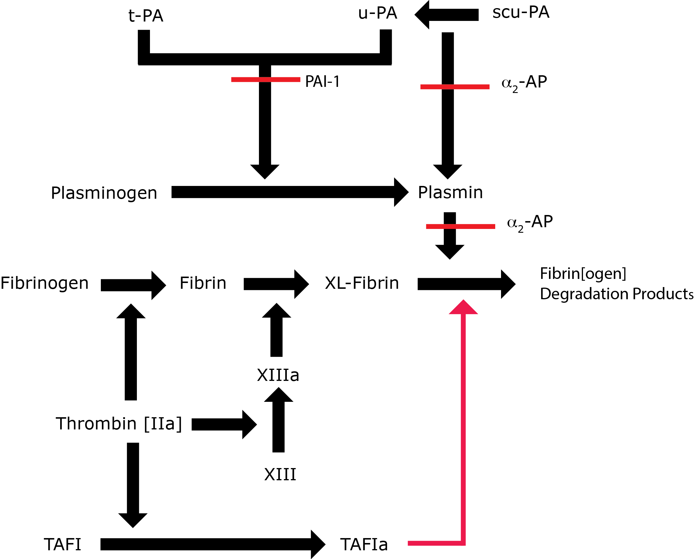
Key: t-PA - Tissue Plasminogen Activator; u-PA - Urokinase-type Plasminogen Activator; XL-Fibrin - cross-linked fibrin; TAFI - Thrombin Activatable Fibrinolytic Inhibitor; TAFIa - Activated Thrombin Activatable Fibrinolytic Inhibitor; α2-AP - Alpha-2-antiplasmin [Alpha2-Plasmin Inhibitor]; scu-PA - single chain Urokinase-type Plasminogen Activator. A red line indicates an inhibitory pathway.
Principles & Methodology
A number of methods are available for measuring u-PA both immunologically and functionally:
1. Immunological ELISA assays: These generally involve coating of a monoclonal antibody specific for u-PA onto a microtitre plate. Plasma samples and standards are added to the plate an incubated. The u-PA binds to the mouse monoclonal antibody and after washing a biotinylated polyclonal antibody specific for u-PA is added and after washing the bound antibody is detected using an Avidin-Biotin-Peroxidase Complex. The change in colour is directly proportional to the u-PA concentration.
2. Functional assays:
a. Chromogenic Assays: The assay measures the ability of u-PA to convert Plasminogen to Plasmin using a Plasmin-specific synthetic substrate. The amount of Plasmin produced is quantitated using a specific Plasmin substrate which on cleavage releases pNA - a chromophore. The change in absorbance 405 nm is recorded and is directly proportional to u-PA activity.
b. Flourometic Assays: These use a peptide substrate that contains the recognition sequence for u-PA. u-PA present in the sample catalyses the cleavage of the substrate and releases AMC [7-Amino-4-Methyl Coumarin], which is quantified by measuring its fluorescence.
Interpretation of u-PA assays
In the case of u-PA assays and their significance and in part due to the expanding knowledge in this area, review of the current literature is essential.
Reference Ranges
u-PA antigen concentrations range from 2 to 7 ng/ml. Increased levels may be seen in patients with liver disease.
What Test Next?
In cases of suspected disordered Fibrinolysis, the TEG-ROTEM can be very useful as a global assessment of Fibrinolysis. Sequence analysis of the relevant genes is also fundamental to the investigation of someone with suspected disordered Fibrinolysis.