Introduction
These tests use monoclonal antibodies specific for the platelet GpIb binding site of VWF located in the A1 domain of the molecule and are independent of Ristocetin.
A number of Epitope-specific assays are available and these are summarised below:
| Abbreviation | Description |
|---|---|
| VWF:GPIbR | An assay that is based upon the Ristocetin-induced binding of VWF to a recombinant wild-type GPIb fragment |
| VWF:GPIbM | An assay that is based upon the spontaneous binding of a gain-of-function mutant GPIbα fragment |
Principles & Method - see also VWF:RCo Assays
1. ELISA assays: In this assay microtitre plates are coated with a monoclonal antibody directed against part of the VWF factor that binds to the platelet GpIb receptor. Plasma samples [reference, tests and standards] are diluted and aliquots added to the wells of the microtitre plate. The plates are incubated during which the immobilised antibody binds to the VWF. After incubation the bound VWF is detected with an anti-human horseradish peroxidase [HRP] labelled antibody. The bound antibody is quantified in a colorimetric reaction by adding the HRP substrate OPD [1,2 ortho-phenylendiamine dihydrochloride], and measuring the absorbance at 492 nm. The absorbance is proportional to the concentration of VWF. A calibration curve is constructed by plotting on VWF:Act against OD on linear-linear graph paper and from which the concentration of the test samples can be derived [see reference 2]. This test is independent of ristocetin.
2. Automated latex agglutination method: This
utilises latex particles coated with a monoclonal antibody directed against the part of VWF that binds to the platelet GpIb receptor. The activity of VWF is determined by measuring the increase of turbidity produced by the agglutination of the latex particles as a consequence of the interaction between the GpIb receptor of VWF and the monoclonal antibody. This test is independent of ristocetin, can be fully automated and run on a number of analysers.
Interpretation
1. Using an ELISA assay, in patients with Type 1 or 2A VWD, the results correlate with VWF:RCo activity. In patients with Type 1 VWD, VWF:Act measured by the monoclonal ELISA are similar to levels of VWF:Ag measured in a polyclonal antibody-based ELISA. The VWF:Act results are significantly lower than VWF:Ag in Type 2A and 2B plasma allowing the discrimination of variant VWD. In 2M VWD the VWF:Act is borderline to low. Individuals with Type 3 VWD will show very low or undetectable VWF:Act using the VWF:Act assay.
2. The Latex Agglutination technique uses a similar antibody and the interpretation of the test is similar.
Reference Ranges
The reference range for VWF:Act assays are usually in the region of 50-150 IU/dL.
Von Willebrand factor is an acute phase protein and so the levels may rise at times of stress e.g. in pregnancy, following surgery.
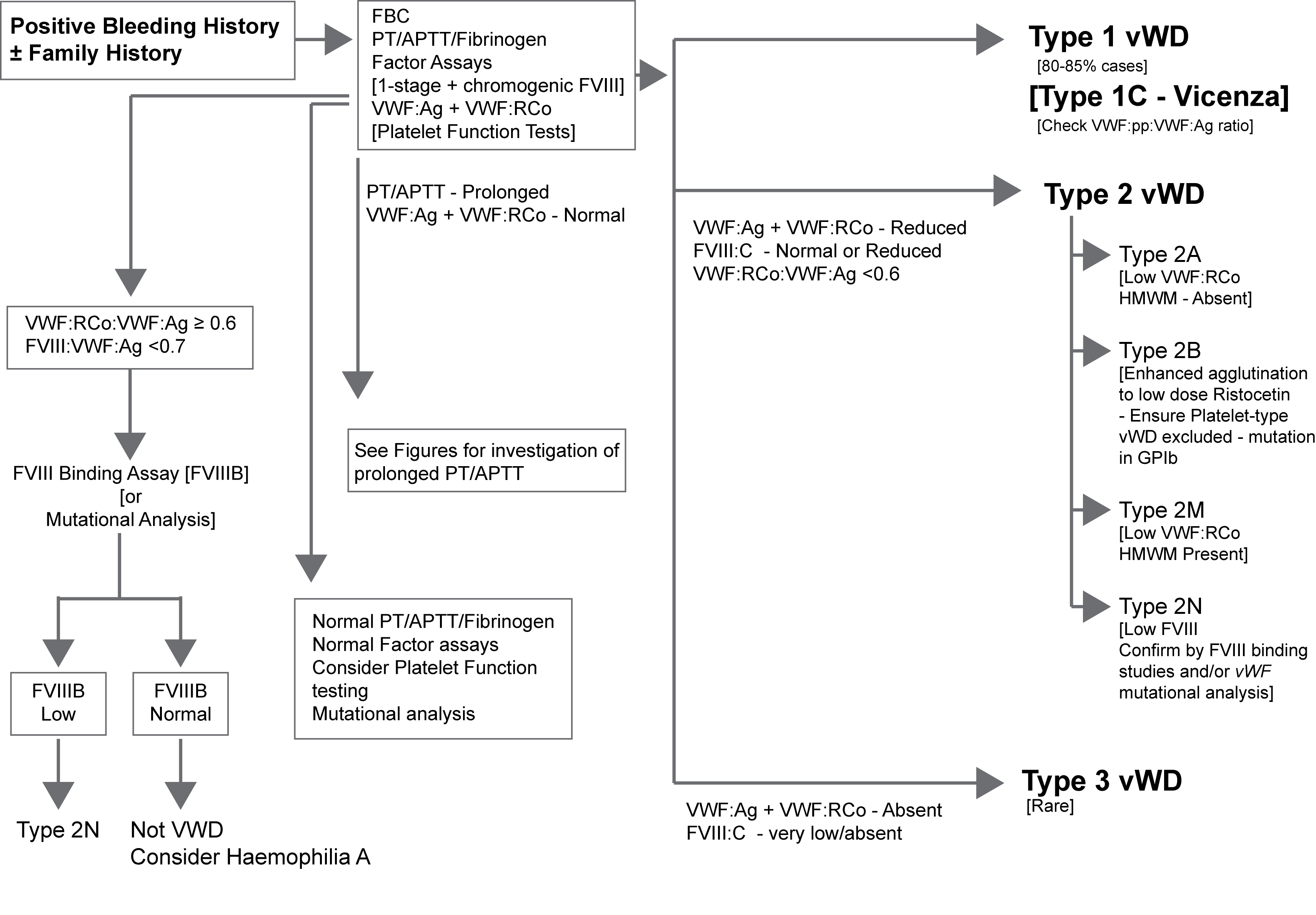
What Test Next
Measurement of VWF activity is usually undertaken as part of a screen to establish or exclude a diagnosis of von Willebrand disease. These assays are also used to monitor replacement therapy in individuals with VWD.
The illustration below outlines one approach to the diagnosis of suspected VWD. Click HERE for an expanded version of this illustration.